


 View Map
 Book an Appointment
 Find a Doctor
 Health Check-up


How Brain Imaging Helps Doctors Decide the Right Stroke Treatment
Mar 18, 2026
Category: Blog
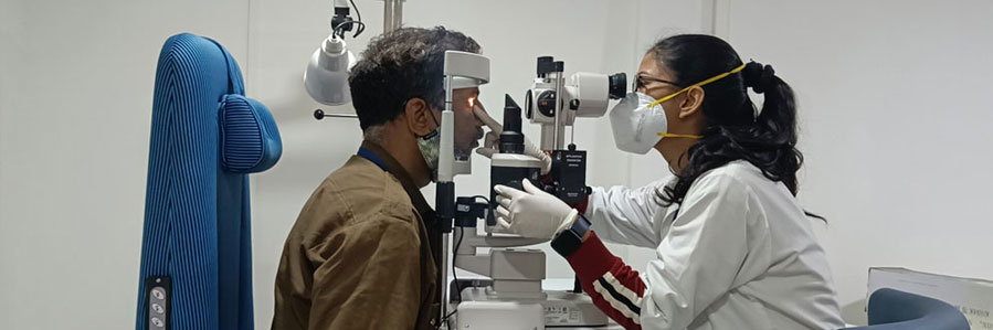
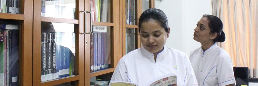
At Jupiter Hospital, we are equipped with over 30 specialty treatments. There are specialised departments dedicated to illnesses which are backed by skilled and experienced doctors and team of healthcare professionals who are also experts at their craft.
Have a query or need to visit an expert? Book an appointment online to consult our doctors and we’ll take care of your needs.
Established in 2007, Jupiter Hospital is a tertiary care Hospital with a ‘Patient first’ ideology deeply instilled in its foundation, to deliver leading-edge healthcare to cater to the changing needs of the growing populace.


Through Jupiter Foundation, we ensure world class health care for the people that are economically challenged. Jupiter Foundation works on the philosophy of putting the patient first in everything.
At Jupiter, we believe there is a better path to healing that humanizes the practice of health care and inspires hope in the Patient & family who need it most. Our primary value – The needs of the patient come first – is at centre of our plans and decisions. You'll also find that our pride – in where we work, and in what we do – is a common trait. You will be truly part of an amazing team committed to solving the most serious and complex medical challenges.


Eastern Express Highway, Service Rd, Next To Viviana Mall, Thane,
Maharashtra - 400601
Jupiter Hospital, Baner,
Pune.
Scheme No. 94, Sector 1,Ring Road, Near Teen Imli Square,
Indore,
Madhya Pradesh - 452020
Jupiter Hospital, Kalyan Shill Road,
Dombivli 421204

Mar 18, 2026
Category: Blog
By Dr. Chaitanya Pauranik, HOD Radiology and Chief Radiologist, Vishesh Jupiter Hospital, Indore
In stroke care, every minute counts. Nearly 1.9 million brain cells are lost per minute when blood flow to the brain is blocked. This is why clinicians often say time is brain. But speed alone is not enough. Precision is equally critical.
Before any stroke treatment begins, doctors must answer four key questions. Is the stroke caused by a clot or a bleed? Is brain tissue still salvageable? Is a major artery blocked? Is aggressive treatment safe?
The only way to answer these questions reliably is through rapid, structured brain imaging. Modern stroke management is imaging-driven and protocol-based. Imaging does not just confirm the diagnosis. It determines whether a patient receives clot-busting medication, undergoes mechanical thrombectomy, or requires neurosurgical management.
Understanding how different imaging modalities guide stroke treatment helps patients and families appreciate why immediate scanning is the first and most important step in emergency stroke care.
| Imaging Modality | Primary Role | Treatment Impact |
|---|---|---|
| Non-Contrast CT (NCCT) | Rule out hemorrhage | Determines eligibility for thrombolysis |
| CT Angiography (CTA) | Detect large vessel occlusion | Identifies thrombectomy candidates |
| CT Perfusion (CTP) | Assess infarct core vs penumbra | Extends treatment window in selected patients |
| MRI Brain | Detect early infarct, stroke age | Longer hospital stay |
Advanced imaging can extend the mechanical thrombectomy window up to 24 hours in carefully selected patients.
The first imaging test in suspected stroke is usually a Non-Contrast CT scan. It is fast, widely available, and highly effective in detecting intracranial hemorrhage.
Key findings include:
If bleeding is detected, the treatment pathway shifts immediately toward blood pressure control and neurosurgical consultation.
CT Angiography – Detecting Large Vessel Blockage
CT Angiography visualises blood vessels in the brain. It helps identify major artery blockages such as internal carotid artery, middle cerebral artery, or basilar artery occlusion.
If a large vessel occlusion is confirmed, the patient may be eligible for mechanical thrombectomy, a procedure that physically removes the clot.
CT Perfusion – Identifying Salvageable Brain Tissue
CT Perfusion differentiates between
Infarct core – Irreversibly damaged tissue
Penumbra – Hypoperfused but salvageable brain tissue
If imaging shows a small core and large penumbra, aggressive reperfusion therapy can be life-saving. This technology allows treatment decisions beyond traditional time windows.
MRI Brain – Detailed Stroke Assessment
MRI is particularly valuable in
Wake-up strokes
Posterior circulation strokes
Stroke mimics
Key sequences include
DWI for detecting acute infarct within minutes
FLAIR for estimating stroke timing
GRE or SWI for detecting microbleeds
MRA for vessel assessment
MRI provides high-resolution detail when clinical scenarios are complex.
Imaging-Guided Treatment Pathways
Hemorrhagic Stroke
Ischemic Stroke Without Large Vessel Occlusion
If within 4.5 hours- IV thrombolysis Beyond 4.5 hours- Perfusion-based selection may apply Ischemic Stroke With Large Vessel Occlusion CTA confirms occlusion CTP confirms salvageable tissue Mechanical thrombectomy considered Stroke Imaging Algorithm Step 1
Non-Contrast CT to rule out bleed
Step 2
CT Angiography to detect large vessel occlusion
Step 3
CT Perfusion or MRI to assess salvageable tissue
Step 4
Decide between thrombolysis, thrombectomy, or conservative management
Brain imaging in stroke is not merely diagnostic. It directly determines therapy.
Modern stroke care depends on rapid imaging and structured interpretation. The right scan at the right time ensures that treatment decisions are precise, safe, and outcome-driven.
To quickly rule out bleeding and guide urgent treatment decisions.
No. Rapid imaging accelerates appropriate treatment.
Most patients recover within a few days and return to daily activities sooner compared to traditional surgery.
Not always. CT-based protocols are sufficient in many acute cases.
Eligibility depends on cancer stage, overall health, and the doctor’s assessment.
No. Imaging is mandatory before thrombolysis or thrombectomy.
Yes. It helps select the right patients for life-saving interventions.
Stroke treatment today is fast, structured, and imaging-guided. Brain imaging allows clinicians to move beyond guesswork and deliver targeted therapy within the critical golden window.
It offers enhanced precision, reduced bleeding, faster recovery, and better preservation of urinary and sexual function.
Yes. It is a highly controlled and surgeon-guided procedure with proven safety and successful outcomes.
Most patients recover within a few days and return to daily activities sooner compared to traditional surgery.
The advanced precision helps protect surrounding structures, improving urinary control post-surgery.
Eligibility depends on cancer stage, overall health, and the doctor’s assessment.
Patients can visit Vishesh Jupiter Hospital, Indore, for consultation with Dr. Abhishek Laddha, Uro-Oncologist.
Robotic surgery has redefined prostate cancer care, offering new hope for men over 45. By combining cutting-edge technology with surgical expertise, Vishesh Jupiter Hospital ensures superior outcomes, faster recovery, and improved quality of life. If you’re over 45, it’s time to take charge of your prostate health - get screened, stay aware, and consult our experts today.

Blog
Mar 18, 2026
Blog
Mar 18, 2026
Blog
Mar 18, 2026
Blog
Mar 18, 2026
Blog
Mar 18, 2026

Blog
Mar 18, 2026

Blog
Jan 16, 2026
Blog
Apr 13, 2023
Blog
Apr 13, 2023
Blog
Apr 13, 2023
Blog
Apr 13, 2023
Blog
Apr 13, 2023
Blog
Apr 13, 2023
Blog
Apr 13, 2023
Blog
Apr 13, 2023
Blog
Apr 13, 2023
Blog
Apr 13, 2023
Blog
Apr 13, 2023
Blog
Apr 13, 2023
Blog
Apr 13, 2023
Blog
Apr 13, 2023
Blog
Apr 13, 2023
Blog
Apr 13, 2023
Blog
Apr 13, 2023
Blog
Apr 13, 2023
Blog
Apr 11, 2023
Blog
Apr 11, 2023
Blog
Apr 10, 2023
Blog
Apr 10, 2023
Blog
Apr 10, 2023
Blog
Apr 10, 2023
Blog
Apr 10, 2023
Blog
Apr 10, 2023


 Find a Doctor
 Health Checkup
 Book an Appointment